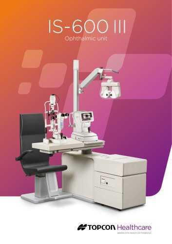
In the realm of modern eye care technology, mastering sophisticated diagnostic tools is crucial for delivering precise and effective patient care. This guide is designed to help you navigate the features and functionalities of your high-end ophthalmic device, providing you with the essential knowledge to optimize its use. From setting up the equipment to understanding its various functions, this resource will ensure you can make the most out of your advanced imaging system.
By familiarizing yourself with the operational procedures and technical details, you’ll be better equipped to perform comprehensive eye examinations with confidence. Our comprehensive overview covers everything from initial setup to advanced features, aiming to streamline your workflow and enhance diagnostic accuracy.
Whether you are a seasoned practitioner or new to this sophisticated technology, the insights provided here will be invaluable in helping you operate your device efficiently. Embrace the opportunity to elevate your practice with a deeper understanding of your equipment’s capabilities and functionalities.
Overview of Topcon TRC NW8
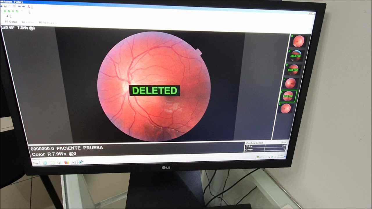
This section provides a comprehensive introduction to a highly advanced piece of ophthalmic imaging equipment. Designed for precision and clarity, this device plays a crucial role in the examination and analysis of the retina and other key structures of the eye. Its state-of-the-art technology allows for detailed imaging, enabling practitioners to diagnose and monitor a wide range of eye conditions with greater accuracy.
The system features advanced capabilities that enhance the quality of imaging, including high-resolution outputs and various adjustable settings to tailor the examination process. The integration of sophisticated components ensures that the device meets the high standards required for professional use in clinical settings. Understanding the functionality and operational aspects of this equipment is essential for leveraging its full potential in patient care and diagnostic procedures.
Understanding the Device Features
Exploring the functionalities of a sophisticated imaging apparatus reveals its broad range of capabilities designed to enhance diagnostic precision and user experience. This section delves into the core attributes of such a device, shedding light on how each feature contributes to its overall performance and usability.
- High-Resolution Imaging: The device is equipped with advanced optics that ensure high-resolution images, allowing for detailed and accurate analysis of the retinal structures.
- Automatic Focus Adjustment: An intelligent autofocus system helps maintain clarity and sharpness in various imaging conditions, reducing the need for manual intervention.
- Wide Field of View: The wide-angle capability offers a comprehensive view of the retina, facilitating the detection of a wider range of conditions with a single capture.
- Enhanced Light Sensitivity: Improved sensitivity to light enables clear imaging even in lower light conditions, ensuring reliable results across different environments.
- Ergonomic Design: The user-friendly design of the device provides comfort and ease of use, promoting efficiency during prolonged examination sessions.
- Data Integration: Seamless integration with digital systems allows for easy data management and analysis, streamlining workflow and record-keeping.
Understanding these features can significantly enhance the effectiveness of your diagnostic practices, ensuring that each examination is conducted with the highest level of accuracy and convenience.
Step-by-Step Setup Instructions
Setting up your equipment requires careful attention to detail to ensure proper functioning and optimal performance. This guide will walk you through the essential steps needed to get everything ready for use. Each step is designed to be clear and concise, helping you avoid common pitfalls and streamline the setup process.
Begin by selecting a suitable location for your device, ensuring it is on a stable surface and free from any obstructions. Next, connect the power supply, making sure all connections are secure and correctly aligned. Proceed by turning on the equipment and following the on-screen prompts or visual indicators to configure initial settings.
Once the basic setup is complete, calibrate the device as instructed to ensure accuracy. This may involve adjusting various settings or performing a series of tests to confirm proper operation. Finally, review the user interface and familiarize yourself with the controls and features to maximize your experience.
By following these organized steps, you’ll ensure that your equipment is set up efficiently and ready for immediate use.
Operating the TRC NW8 Efficiently
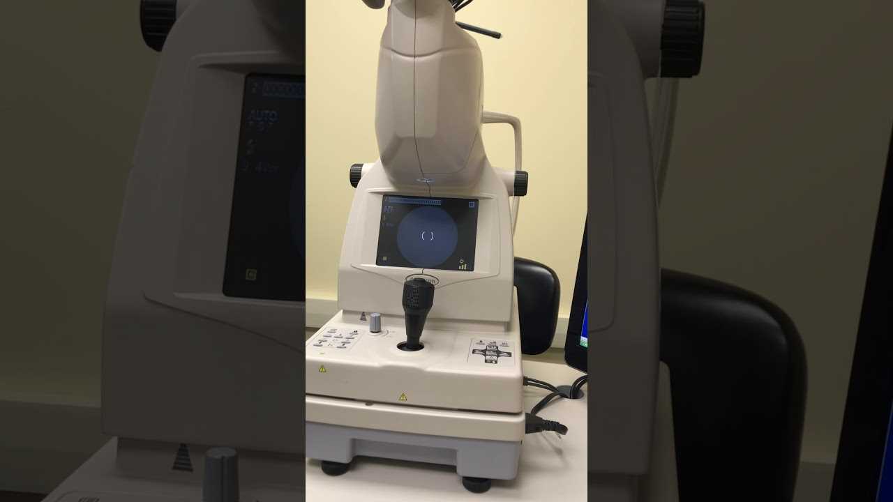
Maximizing the performance of your retinal imaging device requires understanding its optimal use and maintenance practices. Effective operation not only ensures accurate results but also extends the lifespan of the equipment. This section provides essential guidelines to help you operate the device proficiently, enhancing both efficiency and precision in your diagnostic procedures.
To achieve the best outcomes, follow these strategic steps:
| Step | Description |
|---|---|
| 1. Preparation | Ensure that the device is properly calibrated and all settings are configured according to the specific needs of the examination. Regularly check for any updates or maintenance needs to avoid disruptions. |
| 2. Patient Positioning | Correctly position the patient to ensure a clear view and accurate imaging. Adjust the device settings to accommodate different patient sizes and eye conditions for optimal results. |
| 3. Image Capture | Use precise techniques to capture high-quality images. Ensure that the lens and other components are clean and free from obstructions to avoid artifacts and ensure clear imaging. |
| 4. Data Management | Organize and store the images and data efficiently. Implement a systematic approach for data retrieval and analysis to facilitate quick access and accurate diagnosis. |
| 5. Regular Maintenance | Perform routine maintenance checks as outlined in the device’s guidelines. Address any issues promptly to keep the device in top working condition and prevent unexpected failures. |
Adhering to these practices will help you utilize your imaging device to its full potential, ensuring reliable and consistent results in your retinal assessments.
Maintenance and Care Guidelines
Proper upkeep is crucial for ensuring the optimal performance and longevity of optical devices. Regular attention to maintenance tasks not only enhances functionality but also prevents potential issues that could disrupt usage. Adhering to a structured care routine will help maintain the equipment in peak condition and extend its operational lifespan.
Routine Cleaning Procedures
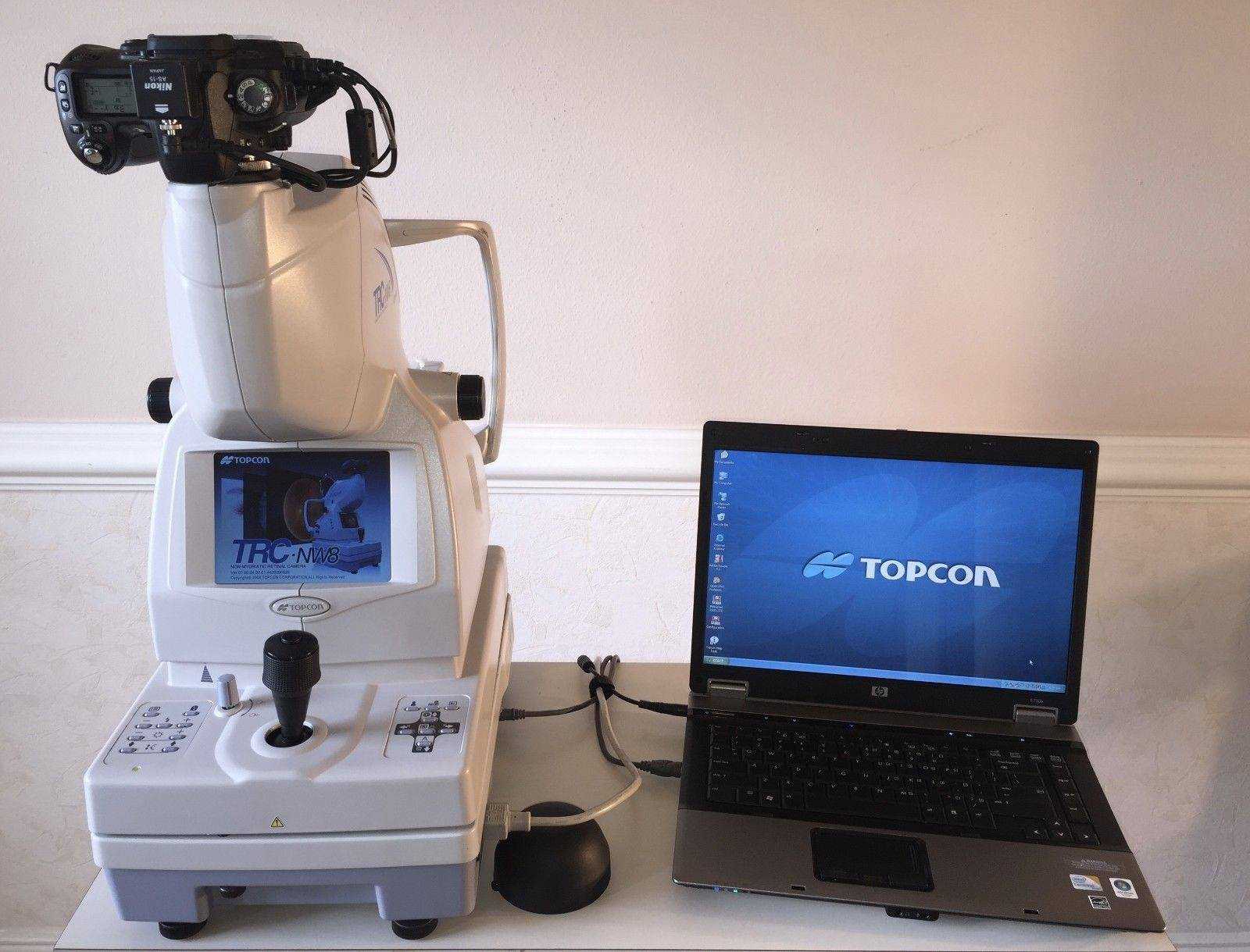
Regular cleaning of optical surfaces and mechanical parts is essential. Use a soft, lint-free cloth to gently wipe the lenses and mirrors, avoiding any abrasive materials that could cause scratches. For more thorough cleaning, use specialized lens cleaning solutions and follow the manufacturer’s recommendations. Ensure that all cleaning is done in a dust-free environment to prevent contaminants from settling on the surfaces.
Periodic Inspections and Servicing
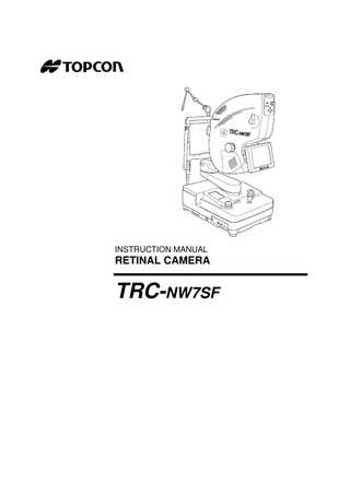
Conduct periodic inspections to check for any signs of wear or malfunction. Regularly verify the alignment and calibration of the device to ensure accuracy in its readings. If any irregularities are detected, consult with a qualified technician for servicing. Keeping a maintenance log can help track any issues and the actions taken to resolve them.
Troubleshooting Common Issues
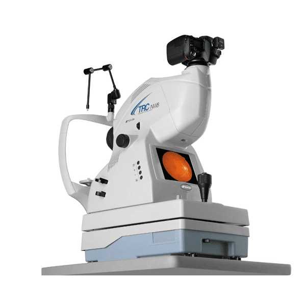
When working with advanced optical equipment, encountering operational problems can be a common occurrence. Understanding how to diagnose and resolve these issues effectively can help ensure smooth and accurate performance. This section provides guidance on addressing frequent challenges users may face, offering practical solutions to keep your device functioning optimally.
| Issue | Possible Causes | Solutions |
|---|---|---|
| Device does not power on | 1. Battery or power source issue 2. Faulty power switch 3. Internal connection problem |
1. Check and replace the battery or power source 2. Inspect and test the power switch 3. Ensure internal connections are secure |
| Image is blurry | 1. Misalignment of optical components 2. Dirty lenses 3. Incorrect settings |
1. Realign optical components according to the guide 2. Clean the lenses with appropriate materials 3. Adjust settings to match the required specifications |
| Unusual noise during operation | 1. Loose internal parts 2. Mechanical malfunction 3. Foreign objects inside the device |
1. Tighten any loose internal components 2. Inspect for mechanical issues and replace parts if needed 3. Remove any foreign objects |
| Inaccurate readings | 1. Calibration issues 2. Software malfunctions 3. Environmental factors |
1. Recalibrate the device as per the calibration procedure 2. Update or reinstall the software 3. Check for environmental influences and correct as necessary |
Tips for Optimal Performance
To achieve the best results with your optical imaging device, following some essential guidelines can significantly enhance its effectiveness and reliability. Implementing these practices ensures accurate readings and prolongs the equipment’s lifespan.
1. Regular Calibration: Ensure that your device is calibrated consistently. Proper calibration is crucial for maintaining precision and consistency in measurements.
2. Routine Maintenance: Perform regular maintenance according to the manufacturer’s recommendations. This includes cleaning optical components and checking for any wear and tear.
3. Proper Handling: Handle the equipment with care to prevent any physical damage. Avoid placing it in environments with extreme temperatures or excessive moisture.
4. Use Quality Accessories: Always use recommended accessories and consumables. Using non-standard parts can affect the performance and accuracy of the device.
5. Software Updates: Keep the device’s software up to date. Software updates often include improvements and fixes that enhance functionality and address potential issues.
6. Training and Familiarity: Ensure that operators are well-trained and familiar with the device’s features and functions. Proper training can lead to more accurate results and efficient operation.
By adhering to these tips, you can ensure that your optical imaging device operates at its full potential, delivering reliable and precise results in your work.